Latest Neurology Articles | Wockhardt Hospitals

ARTICLES ON Neurology

Latest Articles on Neurology

Parkinson's Journey from Neglect to Hope

Rediscovering Life: A Parkinson’s Journey from Neglect to Hope

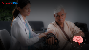
Parkinson’s disease (PD), the second most common neurodegenerative disorder after Alzheimer’s, is often misinterpreted as a natural part of aging. Affecting nearly 1% of individuals over 60, PD frequently goes undiagnosed in its early stages, leading to unnecessary suffering. Dr. Manish Baldia, Functional Neurosurgeon at Wockhardt Hospitals, Mumbai, sheds light on the critical need for early detection and intervention. A retired school principal in his early 70s, exemplifies the challenges of misdiagnosed Parkinson’s disease. For years, he attributed his tremors, slowed movements, and fatigue to aging. His family, though concerned, also dismissed the symptoms as part of the natural aging process. This misconception led to a decline in his quality of life, causing emotional distress and social withdrawal. The turning point came when a visiting relative, a medical professional, urged him to seek specialized care. A neurologist confirmed the diagnosis of Parkinson’s disease, bringing both relief and apprehension. With timely intervention, He was introduced to advanced treatments, including Deep Brain Stimulation (DBS), medication adjustments, and rehabilitation therapy under the expertise of Dr. Baldia. After undergoing DBS, a cutting-edge procedure that regulates abnormal brain activity through electrical impulses, He experienced a dramatic improvement in his motor functions. His tremors reduced, his voice regained strength through speech therapy, and he even resumed hobbies like gardening—activities he once believed were lost forever. A Message of Hope and Awareness Now thriving with his condition, He has become an advocate for Parkinson’s awareness, urging others to recognize early symptoms and seek timely medical attention. “The biggest lesson I’ve learned is not to ignore your body’s signals. Growing older doesn’t mean suffering in silence. Parkinson’s is not the end—it’s a new beginning with the right guidance,” he passionately shares. Dr. Manish Baldia emphasizes, “Early diagnosis and access to advanced treatments can dramatically improve the quality of life for Parkinson’s patients. Our goal is to ensure that no one else has to endure years of unnecessary suffering due to a lack of awareness.” He adds, “With advancements like Deep Brain Stimulation and personalized treatment plans, we can offer hope and a renewed quality of life to those battling Parkinson’s.” Wockhardt Hospitals remains at the forefront of neurological advancements, offering state-of-the-art treatments and compassionate care for Parkinson’s patients. As awareness grows, more individuals can regain control over their lives. _______ Dr. Manish Baldia, Functional Neurosurgeon at Wockhardt Hospitals, Mumbai Central, Shares Insights on “A Parkinson’s Journey from Neglect to Hope” – Exclusive Interview on Zee 24 Taas

Read More
stroke

What Immediate Steps Should We Take if Someone Suffers From a Stroke?

Introduction If you live with someone with a history of stroke or any other ailments that might cause it, it is very important to be aware of how to respond quickly. Since stroke is an acute life-threatening condition, the immediate response goes a long way. Like all medical emergencies, the first and most important thing to do is to call 8108181081 and inform the nearest healthcare centre to assist. Quick response to a stroke can significantly reduce the long-term effects or disabilities that might occur following a stroke. Here is a list of everything you should know about it. What is a Stroke? A sudden disruption in the blood flow to your brain causes a stroke. When your major blood vessels to the brain are affected, brain cells start deteriorating due to the lack of oxygen, which manifests as a stroke. In short, a stroke is the brain’s equivalent of a heart attack. Risk Factors of Stroke Though a stroke can occur in anybody, young or old, they are more common in the later stages of life. Studies show that two-thirds of strokes occur in people older than 65. Apart from age, other medical issues like hypertension, diabetes, and hyperlipidemia also run a higher risk of developing stroke. Any history of stroke and heart conditions is also an important factor here. Stroke remains the second biggest cause of death worldwide and is also a leading cause of disability. Understanding the Signs of a Stroke The following symptoms can present at different intensities depending on the severity of the stroke. Common warning signs of stroke include: Difficulty in maintaining balance. Blurry vision or seeing double. When asking a person to smile, the face might droop to one side. Drooping of the arm when asked to lift straight. Slurred speech is a very common indication. Along with this, one might also have dizziness, vomiting, nausea, neck stiffness, emotional instability, confusion, memory loss, headaches, and in very severe cases even a coma. Things to Do When Someone Has a Stroke It is important to take these measures even if someone has only one of the mentioned symptoms. As said earlier, stroke response at the earliest is life-preserving at best. Here are the crucial steps to follow. Call Emergency Services Whenever you notice the stroke symptoms, call your local emergency service or instruct someone nearby to do so. Stay calm and avoid causing any panic. Get to a Safe Location In case of someone having a stroke in an unsafe or public area like a pool, make sure you take them to a safe and secluded space while waiting for help. Try to position them on their side with a slight head elevation with support. This is to prevent any asphyxia in case they vomit. Check for Breathing You can check this by the airflow through the nostrils or their chest movements. In case of absent breathing, start with chest compressions. Remove any clothing that might be restricting their breathing. Keenly Observe While waiting for help, be careful to look out for any signs of stroke that were mentioned earlier. Remember them carefully to inform the health care provider when the time comes. Communicate Stay calm and reassure those around you. In case the person gains consciousness, make sure not to let them eat or drink anything, as it might pose a high risk of choking. Keep them resting in the same position till help arrives. What Not to Do While someone is having a stroke, make sure you do not allow any of the following things to happen. Always ensure that the person suspected of a stroke receives medical help. Do not dismiss it or let them disregard it as mild discomfort and return to their routine. Even if it is an uncomfortable time of the day, make sure you take that person to the ER. Never let a suspected stroke patient go to the hospital alone. Make sure that you accompany the person or send them with a person trustworthy enough to notice any symptoms and signs that may occur on the way to the hospital. Sending the person to the hospital alone is very dangerous, as the aftermath of a stroke includes loss of consciousness and many other severe issues. Do not allow any self-medication until you reach a healthcare centre and arrive at a proper diagnosis. Even if that is the case, only take medications your treating physician prescribes. Stroke Diagnosis and Treatment Here’s what treatment after stroke looks like: How is the diagnosis made? A physician diagnoses a stroke using neurological examination and imaging. Commonly done tests include a CT scan, lab tests for blood parameters, an ECG or EKG, and MRI scans. Rarely, an EEG might be done to rule out any seizures or similar problems. How is a stroke treated? All patients with stroke are given general advice on controlling their blood pressure and cholesterol levels. Apart from that, specific modes of treatment include thrombolytic drugs and Mechanical Thrombectomy. FAQs Q1. What are the changes I will notice on the face of someone who has had a stroke? The face might droop to one side after a stroke or during a stroke. This becomes more obvious when the patient is asked to smile. Q2. Why should we not self-medicate? Though some advice giving aspirin for an ischemic stroke, it can be very harmful if the person has had a hemorrhagic stroke. Q3. What are the types of stroke I should know about? Stroke can be of two types: Ischemic and hemorrhagic. Blood clots mostly cause ischemic strokes. A few reasons include atherosclerosis, heart defects, and clotting disorders. Some causes of a hemorrhagic stroke include brain aneurysms, brain tumours, and certain diseases affecting the vasculature of the brain.

Read More

Sleep-Related Movement Disorders

Movement disorders during sleep are numerous with varied aetiology. Aetiology presents range from neurological, psychiatry, medical, or sleep disorders. Hence, a detailed and smart evaluation by a Neurologist helps an individual to get the correct diagnosis and appropriate treatment related to aetiology. The importance of evaluation of such conditions lies behind the fact that many such movement disorders disrupt regular sleep which leads to multiple psychological issues like fatigue, anxiety, depression, and medical conditions like uncontrolled hypertension, increased risk of cardiovascular events, etc. Commonly Witnessed Movement Disorders Encountered in Day-To-Day Neurological Practice 1. Restless leg syndrome (RLS) RLS is a movement disorder known to have a fluctuating course with remission rarely seen. It is an irresistible urge to move legs that usually occur during inactivity or at rest and becomes worse in the evening and night 2. Periodic limb movements of sleep (PLMS) PLMS is defined as periodic episodes of repetitive and highly stereotyped limb movements that occur during sleep. 3. Sleep related Bruxism (SLB) SLB is defined as repetitive jaw-muscle activity characterized by clenching or grinding of the teeth and/or by bracing or thrusting of the mandible during sleep.  4. Sleep related hyper motor seizures (SHE) SHE has clinical manifestations which consist of rapid, hyperkinetic movements as well as tonic/dystonic posturing of the limbs usually brief duration with abrupt onset and offset.   5. Sleep related leg cramps (SRLC) SRLC represents painful contractions of the legs during sleep. It is characterized by suddenly occurring, episodic, persistently painful, involuntary contractions of the calf, hamstrings or foot muscles at night.  6. Benign Sleep Myoclonus of Infancy (BSMI)  (BSMI) is a benign condition can occur anywhere between Day 1 of life to 3 years of life it is characterized by twitches or jerks that occur when an infant is drowsy or asleep.  7. Propriospinal Myoclonus at Sleep Onset (PSM) PSM is defined as a movement disorder characterized by a person experiencing sudden jerks of the neck and abdomen both during drowsiness or trying to fall asleep.  8. Sleep-Related Rhythmic Movement Disorder (SRRMD) SRRMD is characterized by repeated rocking, rolling, and banging rhythmic movement disorder, and may also make rhythmic noises such as loud humming. Such rhythmic movements are commonly seen during infancy, and by 5 years old, only 5% of children are still affected. Risk factors include positive family history, infancy, intellectual disability, and associated other sleep disorders. Normal Variants Sleep starts (hypnic jerks): These are present at all ages and characterized by abrupt, startle-like movements that occur as one is drifting off to sleep, sometimes accompanied by a sensation of falling, a dream-like feeling, or a flashing sensation.  Excessive fragmentary myoclonus (EFM)- EFM is considered a benign, nonprogressive condition characterized by the movement of the fingers, toes, or mouth during sleep, with or without corresponding small visible movements(sometimes only evident by electromyography).  Hypnagogic foot tremor – Hypnagogic foot tremor is a rhythmic movement of the feet or toes lasting several minutes and tends to resolve spontaneously over time. It is seen in both adults and children. It occurs around the time of sleep onset with a periodicity of once every one to two seconds involving one or both feet. The patient may perceive a sensation of movement of the feet.  Management for various types of movement disorders related to sleep can be divided into – proper evaluation and correct diagnosis of the type of disorder the individual is suffering from. Though sleep-related movement disorders are clinically diagnosed, investigations required are blood investigations to rule out secondary causes supplemented by EEG, polysomnography, and nerve conduction study as and when required. The establishment of diagnosis is followed by medications for the management of symptoms and treatment of secondary causes leading to such symptoms. When required, the medication should be complemented with non-pharmacological measures as well. Conclusion Movement disorders related to sleep are numerous. More than one movement disorder during sleep can co-exist in the same individual. As these disorders can lead to sleep disturbances followed by psychological consequences, Sleep-related movement disorders need to be properly diagnosed and treated.  For further enquiry please contact  Dr . Sheetal GoyalSr. Consultant NeurologistWockhardt Hospitals, Mumbai Central

Read More

What is Brain Hemorrhage & it’s Treatment?

Subarachnoid haemorrhage (SAH) or ‘brain haemorrhage ” is a very serious life-threatening neuro-surgical emergency condition requiring immediate diagnosis, transfer to a specialised neuro-care centre and intensive (ICU) management. This condition is often seen in both male and female patients, particularly in middle-aged groups. The patient usually experiences a sudden severe headache (worst ever headache in life) followed by loss of consciousness or seizures. The cause of haemorrhage is usually due to the rupture of an aneurysm in the brain. What is an Aneurysm? In some individuals, there is a defect in the walls of the blood vessels of the brain. This balloon-like outpouching from the wall is the weakest point of rupture. It is like a bomb in the brain which on explosion throws a significant amount of blood under pressure causing haemorrhage within the brain. This leads to a sudden increase in pressure in the brain (ICP) and can cause brain death. 30% of such patients with severe haemorrhage die before reaching a hospital. Almost 50% may lose their life after reaching the hospital in the first 24 hours. Diagnosis of SAH CT scan of the brain gives a prompt diagnosis. Details of the aneurysm like its size, location etc can be obtained further by doing a CT angiogram or a Digital subtraction angiography (DSA). Management of an Aneurysmal SAH Essentially involves management of the initial bleed, its complications and sequelae and also preventing further re-bleed. The haemorrhagic blood may disturb the circulation of brain fluid (CSF) causing the ventricles (waterbags) in the brain to increase in size – a condition called hydrocephalus. This often requires a CSF diversion procedure (EVD)involving the placement of a small tube into the ventricle by drilling a burr hole in the skull and draining the CSF externally. Aneurysmal blood can also cause irritation to blood vessels which can contract in size (vasospasm) and reduce the blood supply to the brain. After initial stabilisation, the aneurysm should be secured to prevent a re-bleed. The aneurysm can be closed by either of the two procedures The clipping procedure involves opening of the skull (craniotomy) and direct visualisation, dissection of the aneurysm under a microscope and occluding its neck by applying a titanium clip while coiling is an endovascular procedure and does not involve a craniotomy. Management of an aneurysmal haemorrhage is very complex and challenging. Results depend on the severity or grade of haemorrhage, age, other comorbidities and the neurological condition of the patient at the time of arrival at the hospital. Prompt shifting of such patients to hospitals with neuro-care facilities may result in saving life and better prognosis. Dr Ashwin BorkarConsultant NeurosurgeonWockhardt Hospital

Read More

Benefits of Keyhole Brain Surgery

A revolution in malignant and benign tumors treatment “The first goal of a neurosurgeon is to restore or maintain my patients’ quality of life. Efforts are at the global forefront of advancing these minimally invasive approaches for a wide range of common and uncommon brain and skull base tumors, various vascular conditions such as aneurysms and craniofacial pain syndromes. Keyhole surgery is the concept of safely removing brain & skull base tumors through smaller, more precise openings that minimize collateral damage to the surrounding scalp, brain, blood vessels & nerves. Fortunately, given advances over the last two decades in technology and instrumentation as well improved brain and skull base anatomical understanding, a majority of brain and skull base tumors can now be removed through this surgical approach (Keyhole). Notably, this approach technically demanding, require specialized instrumentation, significant surgical expertise, and are not appropriate for all tumors. Consequently, there remains a role for conventional larger craniotomies. Approaches to perform Keyhole surgeries: Advantages:

Read More
Neuroendocrine Tumour

Neuroendocrine Tumour: Know about its symptoms and treatment

Neuroendocrine Tumour: Know about its symptoms and treatment | PINKVILLA Prof. (Dr.) Mallika Tewari, Senior Consultant & Program Lead Surgical Oncology, Wockhardt Hospital, Mira Road shared in detail about Neuroendocrine Tumour aka NET. Read on to know. Actor Irrfan Khan passed away in Mumbai after battling colon infection. He was diagnosed with a neuroendocrine tumour, 2 years back. Here, we brief you about the neuroendocrine tumour. Read on to know more about this. Neuroendocrine tumours (NETs) are thought to arise from cells throughout the diffuse endocrine system of the human body. They comprise a broad family of tumours that are in the gastrointestinal (GI) tract (i.e. in stomach small intestine, appendix and rectum), lungs and bronchi, thymus, and pancreas.  Less common sites include adrenals, thyroid, parathyroid the pitutary gland (latter in the brain).NETs are uncommon and account for about 0.5% of all newly diagnosed malignancies. Some grow slowly while some tend to grow quickly. Did you know? Some NETs may produce excess hormones (functional NETs). While others do not release hormones or may not release enough to cause symptoms (those are nonfunctional NETs). Symptoms of neuroendocrine tumour One may exhibit symptoms of NETs depending on the location of the tumour and whether it produces excess hormones. The common symptoms are feeling fatigued, unintentional weight loss, and pain owing to a growing tumour. Due to excess hormones (functional tumours), one may also notice signs such as intermittent flushing & diarrhoea, hypertension, hypoglycemia, frequent urination, dizziness, and skin rash. Thus, immediately visit the doctor, in case you exhibit these symptoms. Causes Still, the exact cause of NETs is not known. Most NETs are sporadic and few are hereditary and a part of inherited genetic syndromes e.g. MEN-1; MEN-2 etc. Risk factors  People of all the age groups may suffer from these tumours. Though, it is rarely seen in children. Are you aware? Gastrointestinal conditions like atrophic gastritis can be the culprits.  Treatment The line of treatment will depend on the stage, size and location of the tumour. The treatment may vary from person to person. Your doctor may ask you to go for surgery to get rid of the tumour. Chemotherapy, radiation therapy, hormone therapy, or targeted therapy can also be given.

Read More
Ataxia

What is Ataxia?- A Neurological condition

Ataxia is a degenerative disease of the nervous system. It is caused by damage to the cerebellum, the part of the brain that is responsible for coordinating movement. Another name for this disease is impaired coordination. It can affect people of all ages. Age of symptom-onset can vary widely, from childhood to late-adulthood. Generally it is described as a symptom of incoordination which can be associated with infections, injuries, other diseases, or degenerative changes in the central nervous system. This form of ataxia is different from neurological disease. Ataxia can also be associated with another medical condition, such as head injury, stroke, MS, alcoholism Complications from the disease are serious and oftentimes debilitating. Some types of Ataxia can lead to an early death. What are the symptoms of ataxia? Individuals with Ataxia often require the use of wheelchairs, walkers, and/or scooters to aid in their mobility. How is Ataxia Diagnosed? It is diagnosed using a combination of strategies that may include medical history, family history, and a complete neurological evaluation. Various blood tests may be performed to rule out other disorders. Genetic blood tests are available for some types of hereditary Ataxia. MRI and CT scans of the brain What are the types of ataxia? Sporadic ataxias: This type usually begin in adulthood and have no known family history. Hereditary ataxias: These are caused by a defect in a gene that is present from the start of a person’s life and can be either dominantly inherited or recessively inherited. Which doctor does one consult for diagnosing ataxia? This is a neurological condition therefore it is advised to consult a neurologist to determine the condition. What is the Treatment for Ataxia? The goal of this treatment is to improve the quality of life and requires an individualized approach. It is important to work closely with a neurologist to develop a plan to address symptoms. Speech and language therapy, occupational therapy, and physical therapy are common treatment options. They are sometimes used in conjunction with medication therapy to manage symptoms such as Depression, Dizziness, Vertigo, imbalance coordination, muscle cramps, neuropathy, Stiffness, Spasticity, Rigidity, Dystonia. If you are experiencing any of the following symptoms please consult our neurology department for further evaluation 

Read More
Diagnosis Migraine

Don’t ignore a headache! Consult a Neurologist

“26-year-old Shweta Nanda, software engineer by profession consulted with Dr. Prashant Makhija, Neurologist at, Wockhardt Hospitals in Mumbai for her frequent headaches. She reported that her headaches started at a much younger age and almost 20-30 minutes prior to the headaches she would notice blurring of vision, subsequently she would start having pulsatile headache. She initially consulted an Ophthalmologist who suggested correction of a minor refractive error but as the headaches persisted she consulted a Physician who attributed it to exam related stress. In the last 3-4 yrs., headaches became more frequent and many a times without “visual warning” and lately she started having analgesics on multiple days in a month. Dr. Makhija found out that Shweta’s neurological examination was within normal limits and all her routine blood investigations as well as repeated brain scans were normal. A detail interview with the patient revealed that what started as infrequent “Migraine with aura” was becoming more frequent and many a times without the “visual auras”. This further transformed over time to “Chronic daily headache”. She was asked to maintain a headache dairy and started on Migraine prophylactics and explained that she may have to continue the medications for a reasonable period of time. She showed remarkable response in three weeks and over time her headaches resolved. Shweta not only regained her confidence at work place but her personal life was also back on track.” There are four major types of headache: vascular, muscle contraction (tension), traction, and inflammatory. The most common type of vascular headache is a migraine A migraine is a severe, painful headache that can be preceded or accompanied by sensory warning signs such as flashes of light, blind spots, tingling in the arms and legs, nausea, vomiting, and increased sensitivity to light and sound. The excruciating pain that migraines bring can last for hours or even days. The typical headache of migraine starts on one side of the head usually around the eye or forehead. A migraine occurs when hyperactive nerve cells send impulses to blood vessels. These blood vessels then constrict, resulting in the release of substances that cause severe pain and discomfort. Migraines differ from a typical headache because they last much longer. Migraine is 2 to 3 times more frequent in women than in men. A person suffering from migraines will always like to relax in the dark and quiet place and most of the times relief can be obtained only when vomiting takes place. Many things can trigger a migraine. These include: • Poor coping life skills • Anxiety or Stress – related to exams, Deadline, work related, interpersonal, family etc. • Travel • Lack of food Lack of sleep or sleeping more than usual • Exposure to bright sunlight • Hormonal changes (in women)- premenstrual migraine • Certain food like cheese, banana, curds, coffee Treatment is definitely required and it is best to see a Neurologist to determine the type and cause of headache. Management of Primary headache disorder in general involves a combination of lifestyle modification, judicious use of analgesics and prophylactics medications if the patient is having frequent Migraine headaches. While management of Secondary headache, apart from symptomatic management with analgesics, is directed towards treating the underlying brain disorder. Most of the time, a headache is self- treated and isn’t properly clinically diagnosed. This causes a longer diagnosis period, making treatment difficult. In India, headaches are underestimated, under-recognised and under-treated. To know more about headache and diagnosis please consult our neurology department at Wockhardt Hospital. The Neurosciences department at Wockhardt Hospitals is well-equipped to take care of patients with complex neurological and neurosurgical problems. In particular, our focus areas are stroke, migraine, Parkinson’s disease and other movement disorders, Epilepsy, vascular malformations and brain/spinal cord tumours. Our renowned team of neurologists evaluate all neurological disorders and have been successful in treating various conditions at the hospital

Read More

Brain Tumour & Neuro-Oncology Services

A brain tumour is a mass or growth of abnormal cells developing in the brain. They can be benign or malignant. The tumours can be primary brain tumours originating in the brain itself or in tissues close to it, such as in the brain-covering membranes, cranial nerves, pituitary gland or pineal gland. Sometime tumour can be a secondary lesion from another cancer in the body and spreads to the brain. Brain tumours can grow rapidly and affect well the function of your nervous system. General signs and symptoms include the signs and symptoms of a brain tumour vary greatly and depend on the brain tumour’s size, location and rate of growth. Headaches are very common, they can be new in onset or there can be changes in pattern of headaches .They can be become more frequent and more severe Sometimes there are episodes of unexplained nausea or vomiting. Vision problems, such as blurred vision, double vision or loss of peripheral vision are quiet common. Gradual loss of sensation or movement in an arm or a leg depending on the lesion .Difficulty with balance and speech difficulties can be associated with tumour lesions. Confusion in everyday matters and personality or behaviour changes is also noticed. Seizures, especially in someone who doesn’t have a history of seizures can also occur. Hearing difficulties are sometimes observed. Diagnosis of brain tumours can generally be carried through a neurological examination. Diagnosis can be carries though CT scans, MRI, PET scan and through biopsies. These are generally advised by a neurologist or neurosurgeon. Neuro-Oncosurgery at Wockhardt Hospital: Neuro-oncology is a sub-specialization that deals with brain tumours and spinal cord tumours. These tumours can be both benign (non-cancerous) or malignant (cancerous). Malignant tumours can be divided into primary – that arise from within the brain and spine and secondary tumours – that have spread to brain and spine from other body parts, known as metastatic tumours. All types of brain and spinal cord tumours may produce symptoms that vary depending on the part of the brain and spinal cord involved. With the advancement of technology (advanced microscopes, intraoperative navigation) and better investigation facilities, the morbidity and mortality associated with the brain and spine surgeries has drastically decreased in last two decades. Treatment of benign brain and spinal cord tumours is done by surgical excision, whereas, in case of malignant brain and spinal cord tumours, either chemotherapy or radiation therapy is required, in addition to surgery. Common benign tumours of brain are meningioma, acoustic neuroma, schwannoma and pituitary tumours. Pituitary tumours can be removed through nose with endoscope or microscope without opening the skull. Generally surgery is suggested for the following in case of brain tumours.  Need for surgery: Some surgical techniques include: Craniotomy: Cranioplasty surgery is the most common surgical procedure for excision of brain tumour. An area of the bone is cut out from the skull, the tumour is removed. The whole process is guided by MRI or CT scans either before the surgery or during the surgery. Neuroendoscopy: It also called keyhole brain surgery is done by making small opening in the brain with the aid of endoscope. Neuroendoscopy enables neurosurgeons to access areas of the brain that cannot be reached with traditional surgery and remove the tumour without cutting or harming other parts of the skull. The procedure is performed using an endoscope, a small telescope-like device equipped with a high-resolution video camera and eye piece on the end to allow the neurosurgeon to navigate and access the tumour. For more information on brain tumours symptoms, diagnosis and treatment contact our centres at Wockhardt hospital.

Read More

Second Opinion